
INGENUITY CREATES MILESTONES
OF INTELLIGENT MANUFACTURING
Beijing, China—Ureteral stricture is a common urological condition, the repair and reconstruction of the ureter pose challenges in urological surgery. Some patients continue to suffer from the condition even after undergoing multiple ureteral repair surgeries, leading to physical and psychological distress and a poor quality of life. On October 30th, Director Xuesong Li from Miyun Hospital, Peking University First Hospital, successfully performed a laparoscopic transabdominal left cheek mucosal graft ureteroplasty using the Toumai® surgical robot. This procedure effectively relieved the patient's pain and discomfort. This breakthrough in the application of the Toumai® Robot demonstrates its ability to assist surgeons in performing surgery in confined anatomical spaces, achieving "precise identification, precise dissection, and meticulous suturing," and ultimately achieving refined surgical treatment.


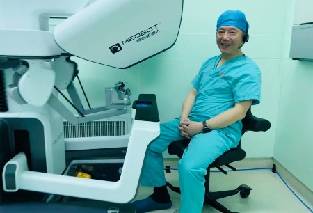
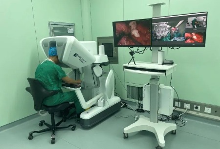
▲The Toumai ® robot-assisted surgery was successfully performed
The patient who underwent this surgery was a 36-year-old woman. She had previously undergone ureteroscopy and stone extraction for left kidney stones at another hospital more than seven months ago. During the procedure, a significant stricture was observed in the upper segment of the left ureter. After placing a double-J stent, the surgery was completed, but the patient experienced recurrent high fever after the stent was removed. She then sought treatment at Peking University Hospital, where Director Xuesong Li diagnosed her with left ureteral stricture and performed a left percutaneous nephrostomy to control the infection. A postoperative CT scan revealed left kidney hydronephrosis, thickening and roughness of the renal pelvis, calyces, and upper segment of the ureter, as well as mild dilation. Antegrade and retrograde combined imaging indicated a significant stricture in the upper segment of the left ureter, leading to a preliminary diagnosis of left ureteral stricture.
To ensure a smooth surgery, preoperative three-dimensional digital medical imaging technology was used to reconstruct the narrow segment, blood vessels, and organs, allowing for visual assessment of the location of the stricture and the surrounding blood supply. Director Xuesong Li and his team analyzed and discussed the surgical plan based on the patient's condition. Considering the complexity of the patient's condition, with a ureteral stricture segment exceeding 2 centimeters, using the patient's own ureteral stricture segment for resection and an end-to-end anastomosis could not guarantee the desired surgical outcome. Additionally, considering the patient's relatively young age, the decision was made to perform a laparoscopic cheek mucosal graft ureteroplasty using the Toumai® Robot to achieve satisfactory treatment results in a single surgery.
During the surgery, with the assistance of Associate Chief Physician Bing Wang and Attending Physician Kunlin Yang, Director Xuesong Li used the Toumai® Robot to access the left colonic gutter, separate intestinal adhesions, and expose the upper segment of the ureter at the lower pole of the kidney. Considering the stricture in the upper segment of the left ureter, he made a longitudinal incision on the anterior wall of the ureterovesical junction and exposed the stricture segment, which was approximately 2.5 centimeters long. The surrounding scar tissue and the stricture segment were resected, and the posterior wall was reinforced with 4-0 sutures to shorten the anastomosis distance. A super slippery guidewire was used to guide the placement of a double-J stent, and a cheek mucosal graft measuring approximately 2.5 * 1.5 centimeters was obtained from the oral cavity and placed in the surgical area to repair the anterior wall defect of the ureter. The graft was sutured with absorbable sutures, and the repaired segment of the ureter was wrapped in the greater omentum. The surgery had minimal blood loss and proceeded smoothly. The patient is currently recovering well, and the long-term efficacy will be further evaluated during follow-up visits.
Research data shows that autologous tissue transplantation for ureteral repair has rapidly developed, and the clinical application of oral mucosal grafts (cheek mucosa/tongue mucosa) for ureteral stricture repair is receiving increasing attention. On the one hand, oral mucosa is smooth and hairless, making it easy to obtain and regenerate. It is located in a warm and moist environment similar to the human body and has thick epithelial tissue, high elastic fiber content, thin lamina propria, and a high density of capillaries, which promotes vascular reconstruction. On the other hand, unlike intestinal mucosa, oral mucosa does not reabsorb metabolic products in urine, avoiding metabolic disturbances in the internal environment. It also does not cause postoperative complications such as urinary tract infections or intestinal obstruction. After transplantation and repair of ureteral stricture using oral mucosal grafts, the transplanted tissue can survive well, and the transplanted site maintains good patency, effectively preventing complications such as graft shrinkage, necrosis, or urine leakage.
Director Xuesong Li stated that the urological upper urinary tract repair team at Peking University has successfully performed over 130 cases of ureteral repair with oral mucosal grafts at both the central and Miyun hospital campuses, with the majority of cases completed using robotic systems. The overall surgical outcomes have been excellent. The key to the robot-assisted laparoscopic cheek mucosal graft ureteroplasty lies in preserving the integrity of the ureteral outer membrane and sheath. The success of the surgery is closely related to the growth and recovery of the reconstructed anastomosis site. Therefore, the segment with the stricture should retain the "ureteral bed" containing the blood supply function as much as possible, as the autologous tissue and the "ureteral bed" have a better chance of survival. Another key point is the precise and stable assistance provided by the surgical robot. During the surgery, the Toumai® Robot demonstrated significant advantages in precise and complex reconstructive procedures. With its immersive 3D high-definition view, magnified 10 times, and flexible wristed instruments, it extended the surgeon's eyes and arms, enabling precise dissection and suturing, greatly improving the surgical precision, and allowing patients to experience less trauma, faster recovery, and significantly improved surgical outcomes.
Mr. Yu Liu, Executive Vice President and Chief Commercial Officer of the MicroPort® MedBot®, stated:
Director Xuesong Li's exceptional and skilled surgical technique, along with the precise and stable assistance of the Toumai® Robot, has successfully expanded the application of this new surgical technique. On behalf of Toumai®, I express sincere gratitude to all the experts involved. In the future, with the guidance and assistance of these experts, we hope to continue expanding the application of the Toumai® in minimally invasive surgeries for various diseases, providing treatment for more patients, improving their quality of life, and realizing the vision of " Make surgery easier, safer and less invasive."


@Copyright 1998-2023,Shanghai Microport Medbot(Group)Co.,Ltd. All rights reserved.
Internet Drug Information Service Qualification Certificate No.: (Hu) - Non-operating -2021-0020
The registered trademarks of “微创", "MicroPort" and"  " are registered trademarks of Shanghai MicroPort Medical (Group) Co., Ltd.” . They have been authorized to be used by Shanghai Microport Medbot (Group) Co., Ltd., and no other party shall use such trademarks without prior written permission thereof.